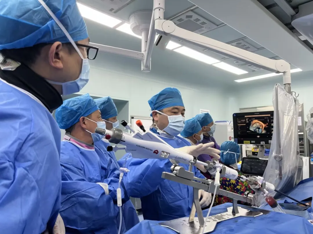
Recently, the cardiology team of Foshan Chancheng Hospital — led by Prof. Peng Yucheng and Prof. Han Wei, with Prof. Zhang Xiaocong, and assisted by Prof. Zhuang Xiaodong and Prof. Yao Fengjuan of the First Affiliated Hospital of Sun Yat-sen University — applied the DragonFly™ transcatheter mitral clip system for the first time and successfully performed a transcatheter edge-to-edge mitral valve repair (TEER) for a patient with severe mitral regurgitation. The smooth completion of the procedure marks another important breakthrough in the team's structural heart intervention capabilities.
Patient Story: A Complex, High-Risk Case
The patient is an 82-year-old man with a complex medical history, including acute coronary syndrome, cardiogenic shock, left heart failure with acute pulmonary edema, sequelae of cerebral infarction, and chronic renal insufficiency. Preoperative echocardiography showed mitral valve prolapse with extremely severe regurgitation, reduced left ventricular systolic and diastolic function, and mild tricuspid and aortic valve regurgitation. He was admitted with a diagnosis of mitral regurgitation, stage 2 hypertension, and atrial fibrillation.
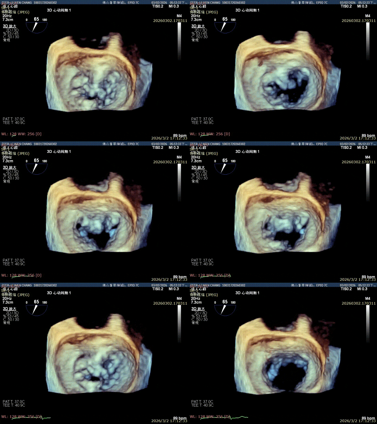
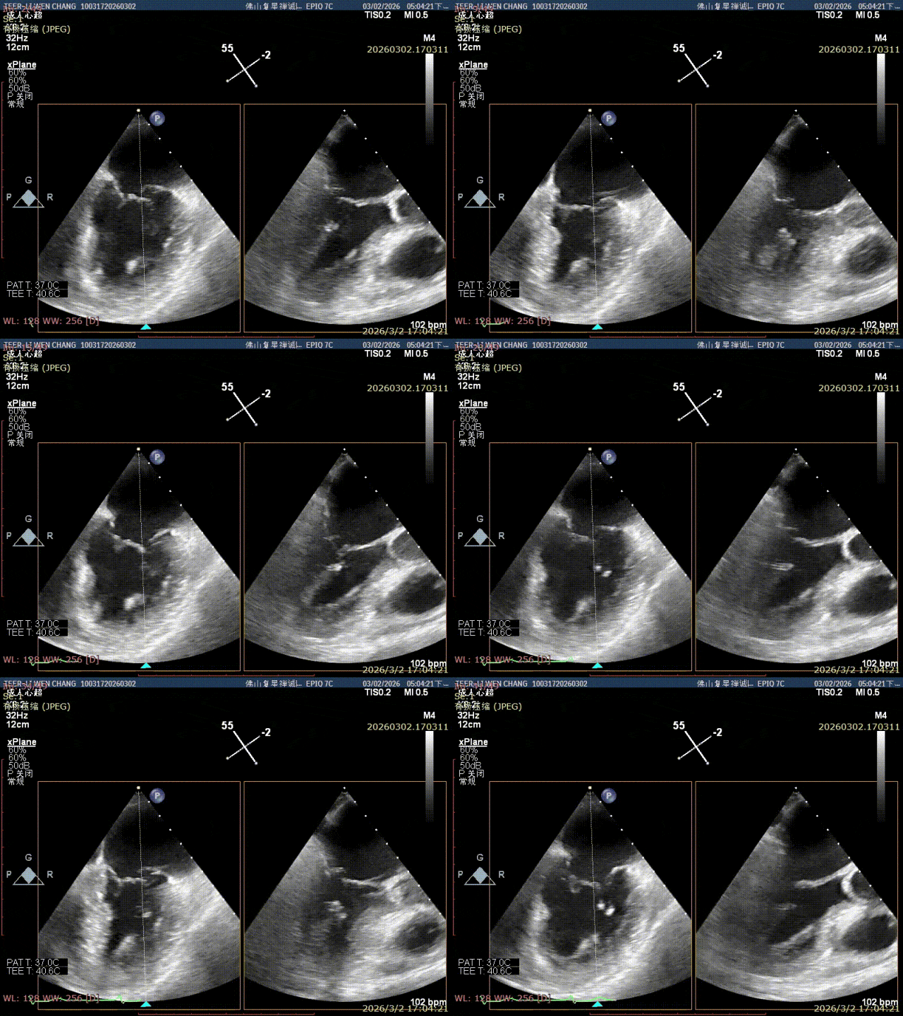
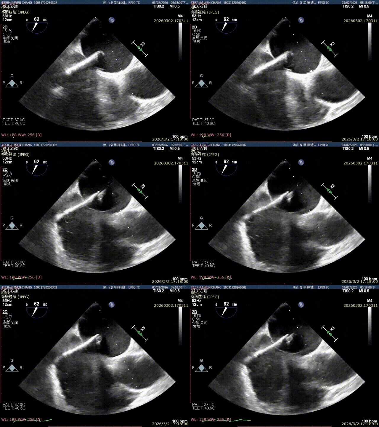
Surgical Challenges and Strategy
Two features made this case demanding. First, the diseased segment sat in the P1 region of the posterior leaflet, where rich chordal structures beneath the valve leave limited working space and demand exceptional precision for positioning and leaflet capture. Second, the lesion was extensive with severe regurgitation, meaning multiple clips might need to be implanted during the same procedure.
After thorough discussion and analysis, Prof. Peng's team decided to use the favorable anatomy of the P1 segment and the appropriate leaflet length: implant one XW0612 DragonFly clip at the P1 region first, then decide in real time — based on the transvalvular gradient and residual regurgitation — whether to add a second clip.
Inside the Procedure, Step by Step
The team performed transseptal puncture (height 4.3 cm), then advanced the guiding sheath and the XW0612 clip. Under real-time imaging they fine-tuned the clip's position, orientation and trajectory, crossed the valve, opened the arms and captured both leaflets, confirmed the leaflets lay flat on the arms, then locked the grippers and gradually closed the clip.
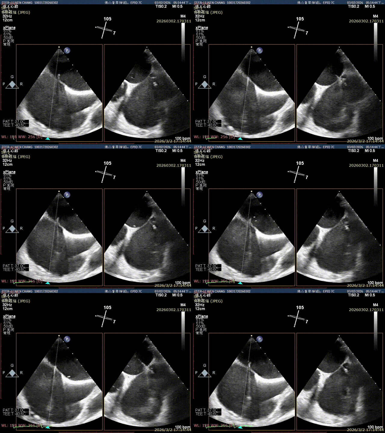
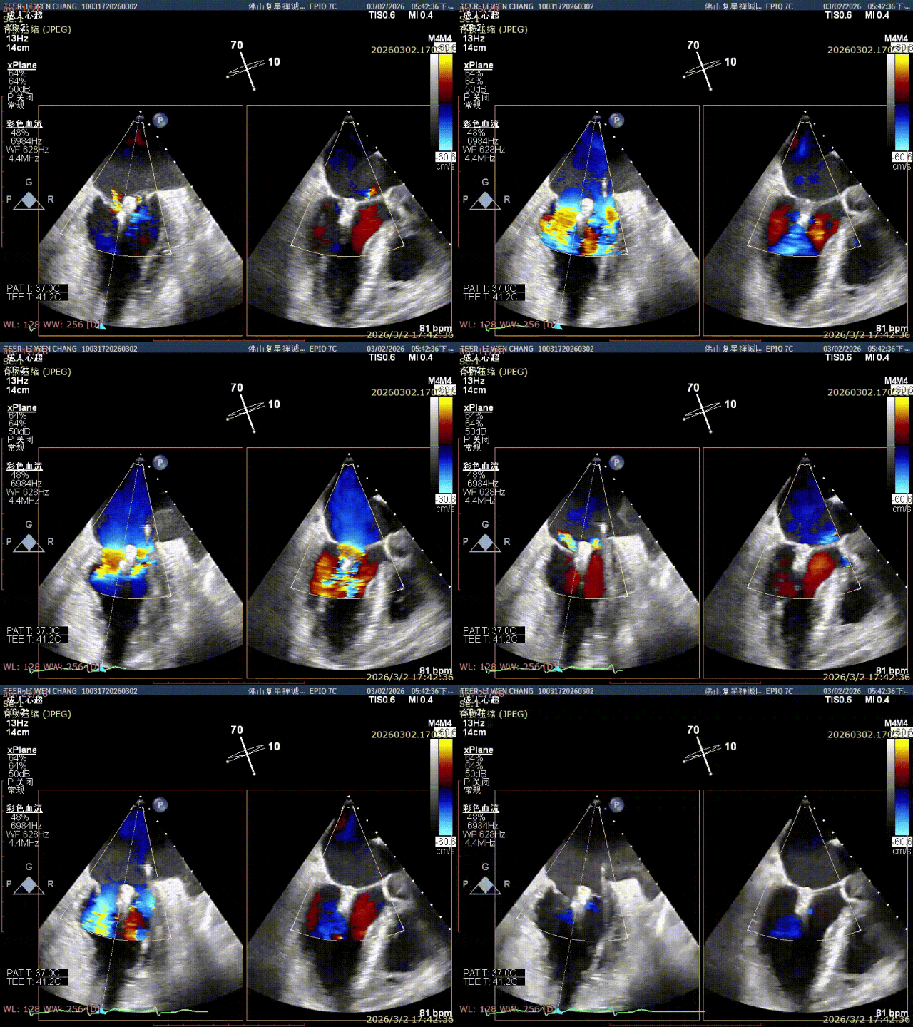
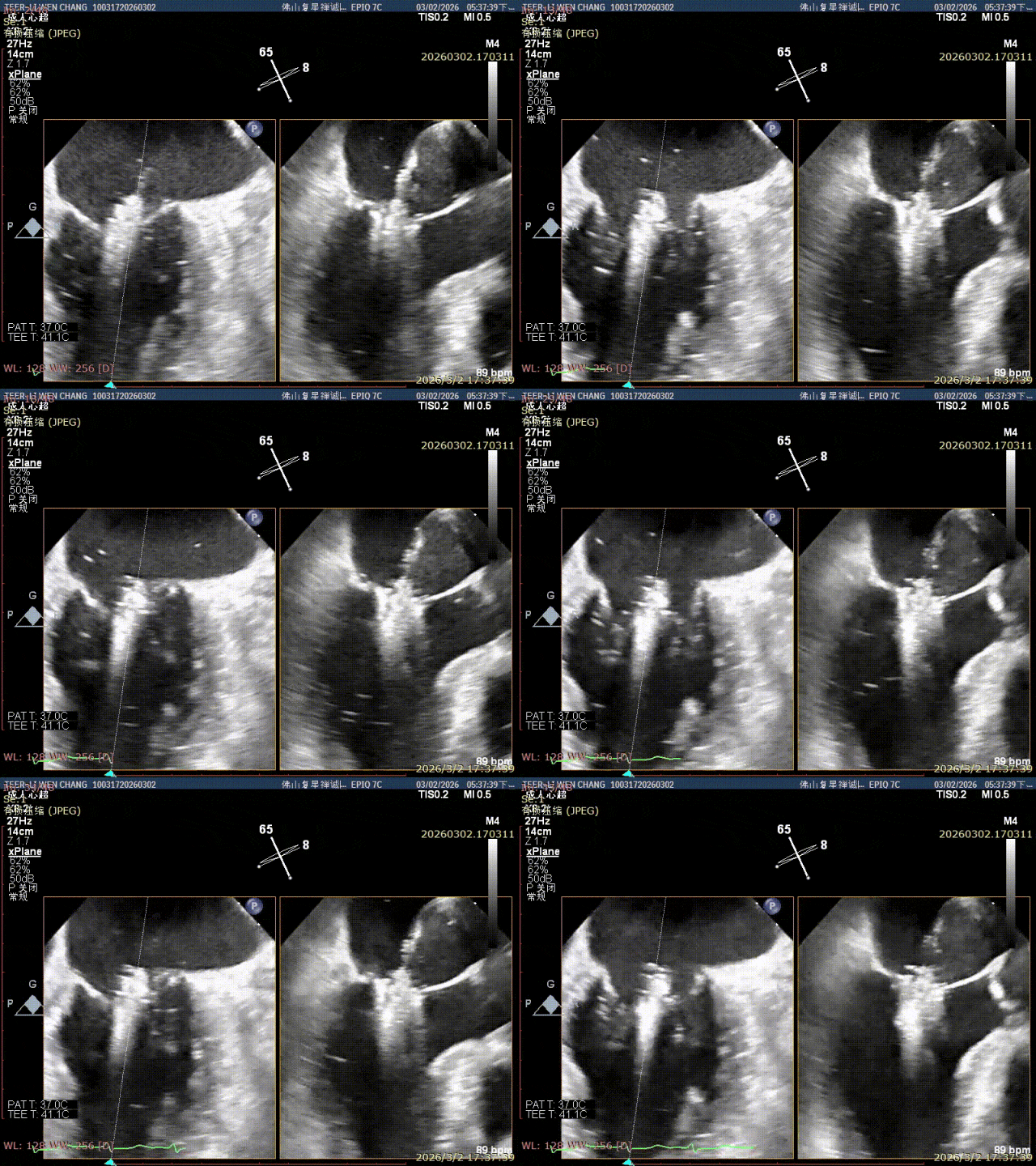
Assessment before and after release showed that trace residual regurgitation remained on both the inner and outer sides of the first clip, and the P1 prolapse persisted — so the team added a second, SN0409 clip lateral to the first. After the second clip captured the leaflets and closed, residual regurgitation disappeared.
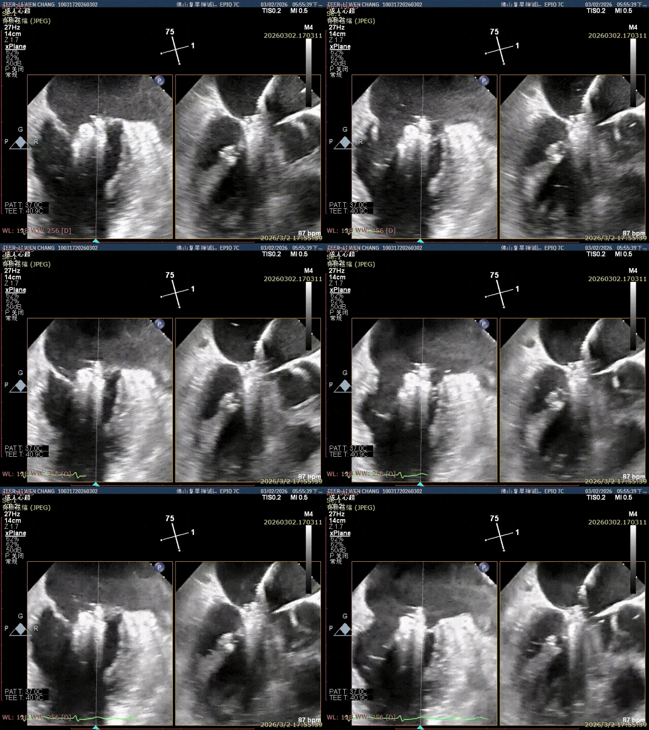
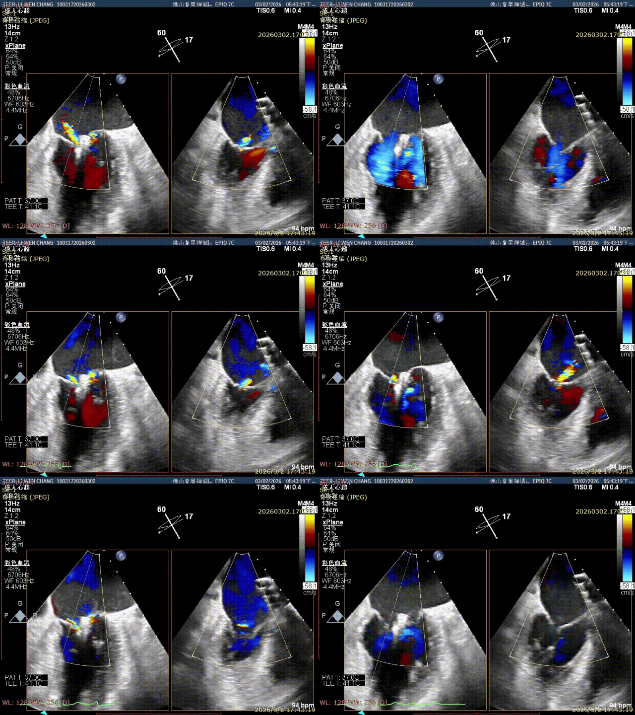
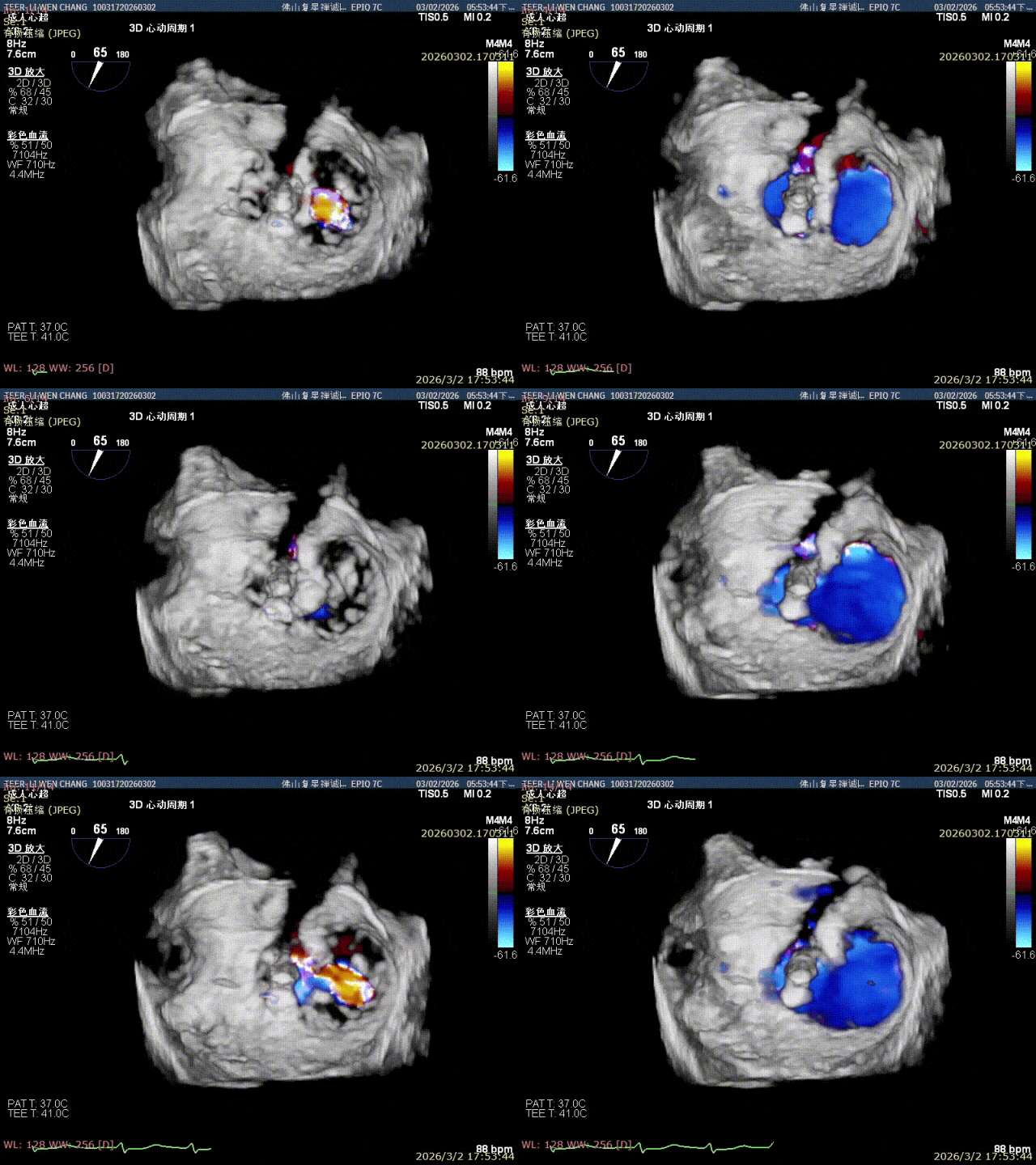
Excellent Immediate Result
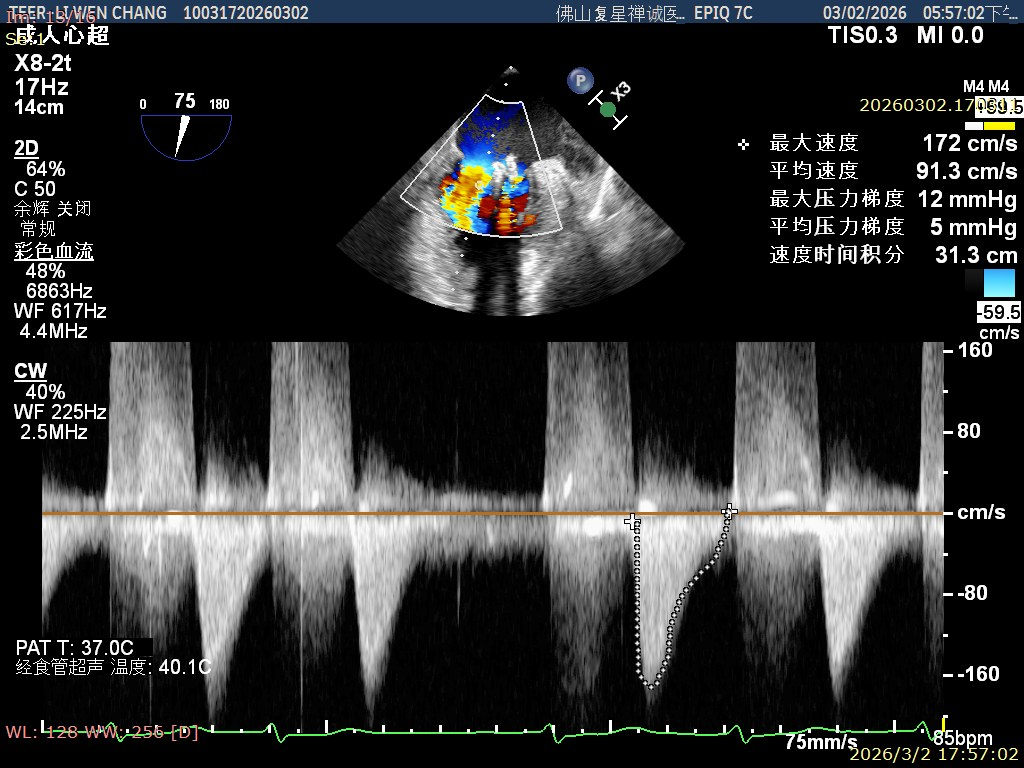
Immediate post-procedure assessment showed the severe mitral regurgitation was dramatically controlled, the mean transvalvular gradient was about 5 mmHg, and the pulmonary vein flow reversal pattern had clearly improved — an excellent hemodynamic result.
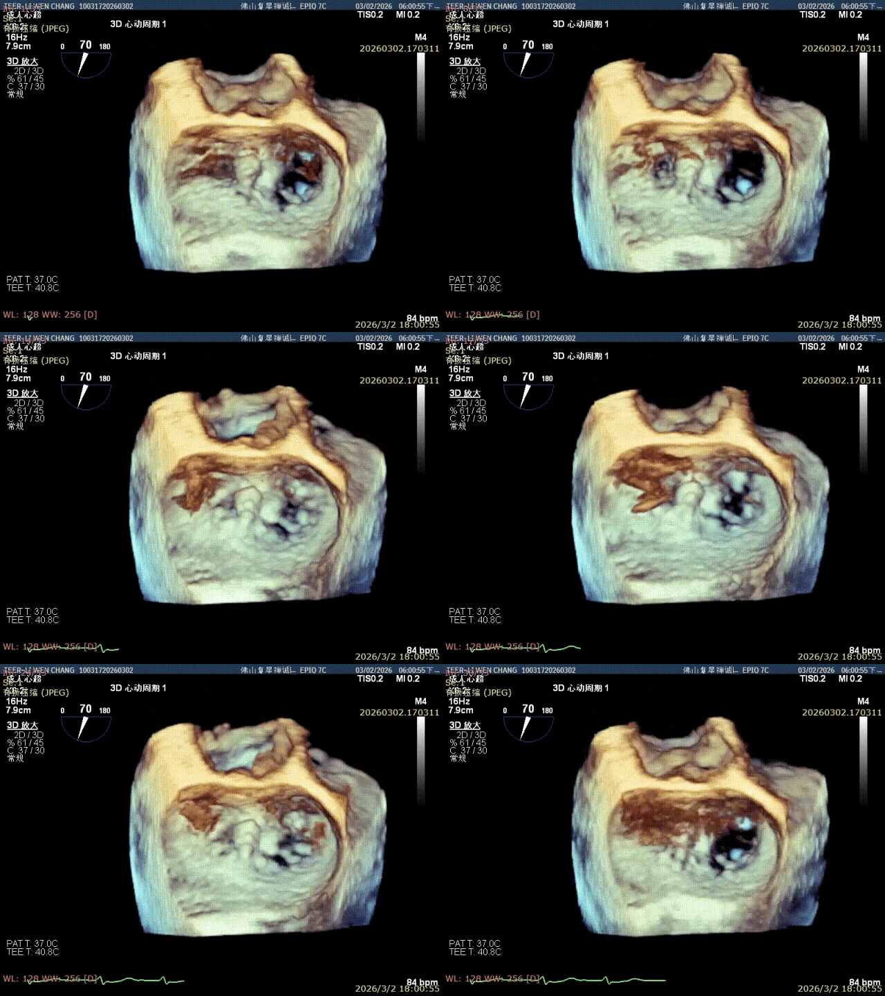
"This patient with severe mitral regurgitation was elderly, with many underlying conditions and complex cardiac anatomy — a case that places extremely high demands on the operator's experience, device-handling skill and intraoperative decision-making," Prof. Peng Yucheng summarized after the procedure. "Backed by a standardized interventional workflow, precise echocardiographic assessment and a flexible surgical strategy, our team completed the dual-clip implantation smoothly. The success of this procedure is another important milestone in minimally invasive structural heart intervention at our hospital. It consolidates the clinical foundation of transcatheter mitral repair in our region, and offers a safe, feasible and efficient new treatment option for more elderly, high-risk patients with severe mitral regurgitation who cannot tolerate conventional open surgery."
About TEER and the DragonFly System
- TEER (transcatheter edge-to-edge repair) clips the leaking mitral valve leaflets together through a vein — no open chest, no cardiopulmonary bypass
- DragonFly™ is China's first domestically developed mitral clip system with both Chinese NMPA approval and EU CE marking ("China-EU dual certification")
- Ideal for elderly or high-risk patients with degenerative (primary) mitral regurgitation who are not candidates for open surgery
Structural Heart Care at Foshan Chancheng Hospital
From TAVR and MitraClip to now the DragonFly TEER system, the Heart Hospital of Foshan Chancheng Hospital continues to expand its structural heart intervention program. Together with the Foshan International Medical Center (FIMC), which serves patients from 100+ countries, elderly and high-risk valve patients — at home and abroad — can access the latest minimally invasive treatment options with a full multidisciplinary team behind every case.